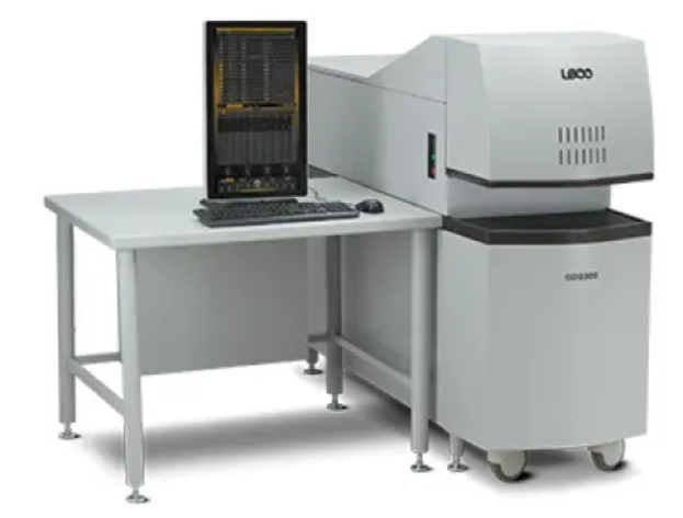
GDS900 Glow Discharge Spectrometer
Glow Discharge Atomic Emission Spectrometer
Our GDS900 Glow Discharge Spectrometer (GDS) provides you with cutting-edge technology for routine elemental measurement in most conductive solid matrices. The platform now includes user-friendly Cornerstone® brand software for improved use, simplified reporting, and faster analysis times, saving you time in the lab.

Features
- When compared to other sources, the calibrations are simple and linear.
- Excitation that takes place away from the sample surface.
- Consumption of reference materials is reduced.
- Stability, adaptability, and performance are all ensured by the detection system.
- From 160 nm to 460 nm, the wavelength range is covered completely.
- Even the most complicated aspects of bulk spectra can be distinguished with a resolution of 50 pm (0.050 nm).
Cleaning between samples automatically saves time and reduces matrix effects, allowing for greater precision.
NANOPIX
Rigaku NANOPIX SAXS/WAXS measurement system is a new X-ray scattering instrument designed for nano-structure analyses. NANOPIX can be used for both small angle scattering (SAXS) and wide angle scattering (WAXS) measurements, which makes it possible to evaluate multi-scale structures from sub-nanometer to nano-order (0.1 nm to 100 nm). It achieves the highest level of small angle resolution (Qmin to 0.02 nm-1) for a laboratory SAXS instrument.
Small angle X-ray scattering (SAXS)
SAXS is a technique used to study nano-scale structures of atoms or molecules as well as their non-uniformity by measuring the diffuse scattering from unequal electron density areas.
SAXS/WAXS measurements for many applications
NANOPIX SAXS/WAXS measurement system is applicable to a variety of materials, such as: solids, liquids, liquid-crystals, or gels (with ordered and disordered structures). Diverse applications include: nano-particle size distribution analyses, three-dimensional protein molecule structure analyses, identification of molecular assembly or disassembly, and research of advanced materials, such as carbon fiber-reinforced plastics (CFRP).
Ultra high performance SAXS/WAXS design
Rigaku NANOPIX SAXS/WAXS measurement system is configured with a high-brilliance, high-power point focus X-ray source, the OptiSAXS high-performance multilayer mirror, the Clear Pinhole high-performance, low scattering pinhole slits, and the HyPix-3000 high-performance 2D semiconductor detector that enables detecting diffraction and scattering even from anisotropic materials. Optionally, the HyPix-6000 detector is also available for wide angle measurements, offering an expanded detection area by combining two detection modules.
Applications
Steel, iron (including as-cast), aluminium, copper, zinc, nickel, cobalt, tungsten, and titanium materials are all good candidates for the GDS900.
Theory of Operation
Glow Discharge Spectrometry (GDS) is a method for determining the elemental composition of solid materials in a direct manner. The glow discharge source is evacuated and backfilled with argon after a prepared flat sample is mounted on it. Between the sample (cathode) and the lamp’s electrically grounded body, a continuous electric field is applied (anode). The spontaneous creation of a stable, self–sustaining discharge, known as a glow discharge, occurs as a result of these conditions. The power supply regulates the applied current, and the lamp voltage is maintained by regulating the argon pressure. The electric field accelerates inert gas ions produced in the plasma toward the sample as soon as the plasma is started (cathode). Kinetic energy is transmitted from the inert gas ions to the atoms on the sample surface via cathodic sputtering, causing some of these surface atoms to be expelled into the plasma.
The atoms are subjected to inelastic collisions with intense electrons or metastable argon atoms once they are expelled into the plasma. The sputtered atoms get electrically stimulated as a result of the energy transmitted by such collisions. By generating photons, the excited atoms quickly relax to a lower energy state. The electronic state of the atom from which each photon was emitted determines its wavelength. Because each element has a distinct electronic configuration, its spectrochemical signature or emission spectrum can be used to identify it.
The emission signals from the glow discharge are measured using a spectrometer. The entire optical system is purged with argon to ensure that the media within the spectrometer is clear to ultra-violet light (160-460 nm). The entire emission spectrum from 160 to 460 nm is recorded using photosensitive Charge-Coupled Device (CCD) arrays positioned at the focal plane. The spectrum is converted into an electrical signal by the CCD arrays, which is then digitised and processed to remove dark current signal, normalise pixel response, increase dynamic range, and remove pixelation.
Because each element’s quantity of photons released is proportional to its relative concentration in the sample, analyte concentrations can be determined by calibrating with known-composition reference samples.
